

Electrophysiology
Electrophysiology examinations allow to measure the voltage of cells in the retina and the visual system triggered by light stimuli. Thus, the activity of the retina and optic nerve can be measured objectively. These are non-invasive examinations, and the electrodes are placed either on the skin (temple, forehead, skull) or the cornea.
In Ganzfeld electroretinography (Ganzfeld ERG), the sum responses of the entire retina are measured, either dark-adapted (scotopic) or light-adapted (photopic). In dark-adapted conditions, the activity of the approximately 120-130 million rod photoreceptors in a healthy retina is measured; in light-adapted conditions, the 6-7 million cone photoreceptors concentrated in the macula are measured. A filament electrode is placed on the cornea, reference and ground electrodes are taped on temple and forehead. Ganzfeld ERG takes about 90 min, including 30 min of dark adaptation, and is used, for example, to diagnose retinal dystrophies (congenital night blindness, retinitis pigmentosa) and to measure the efficacy of uveitis therapies.
In infants, a simplified protocol can be used (Mini-Ganzfeld ERG).
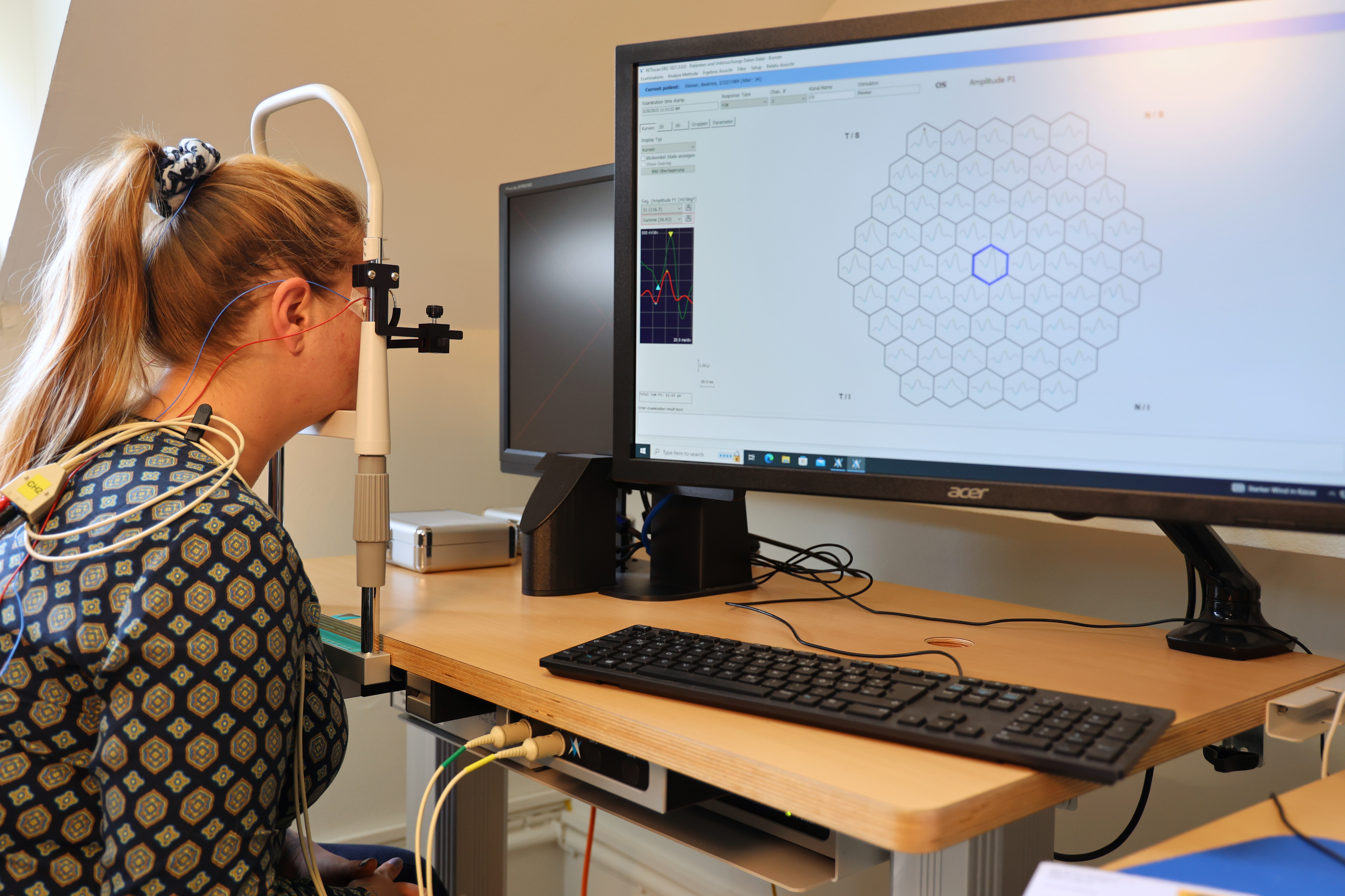
In multifocal electroretinography (multifocal ERG), the center of the retina is stimulated with focal stimuli, providing detailed measurements of macular and peri-macular activity. Since cone photoreceptors are present in the macula, this examination is always performed light-adapted. A filament electrode is placed on the cornea, reference and ground electrodes are taped to the temple and forehead. The multifocal ERG takes about 45 min and is used e.g. for diagnosis of unclear visual loss or retinal dystrophies (macular dystrophies, Morbus Stargardt).
In addition to these routine examinations, the Eye Clinic also offers specific electrophysiology examinations, e.g., the blue-cone ERG (S-cone ERG), pattern ERG, on-off ERG, PNR ERG (photopic negative response), and the electrooculogram.
The blue cone ERG (S-cone ERG) is used to diagnose blue cone hypersensitivity syndrome (Goldmann-Favre syndrome, ESCS) and the electrooculogram (EOG) for the diagnosis of Morbus Best.
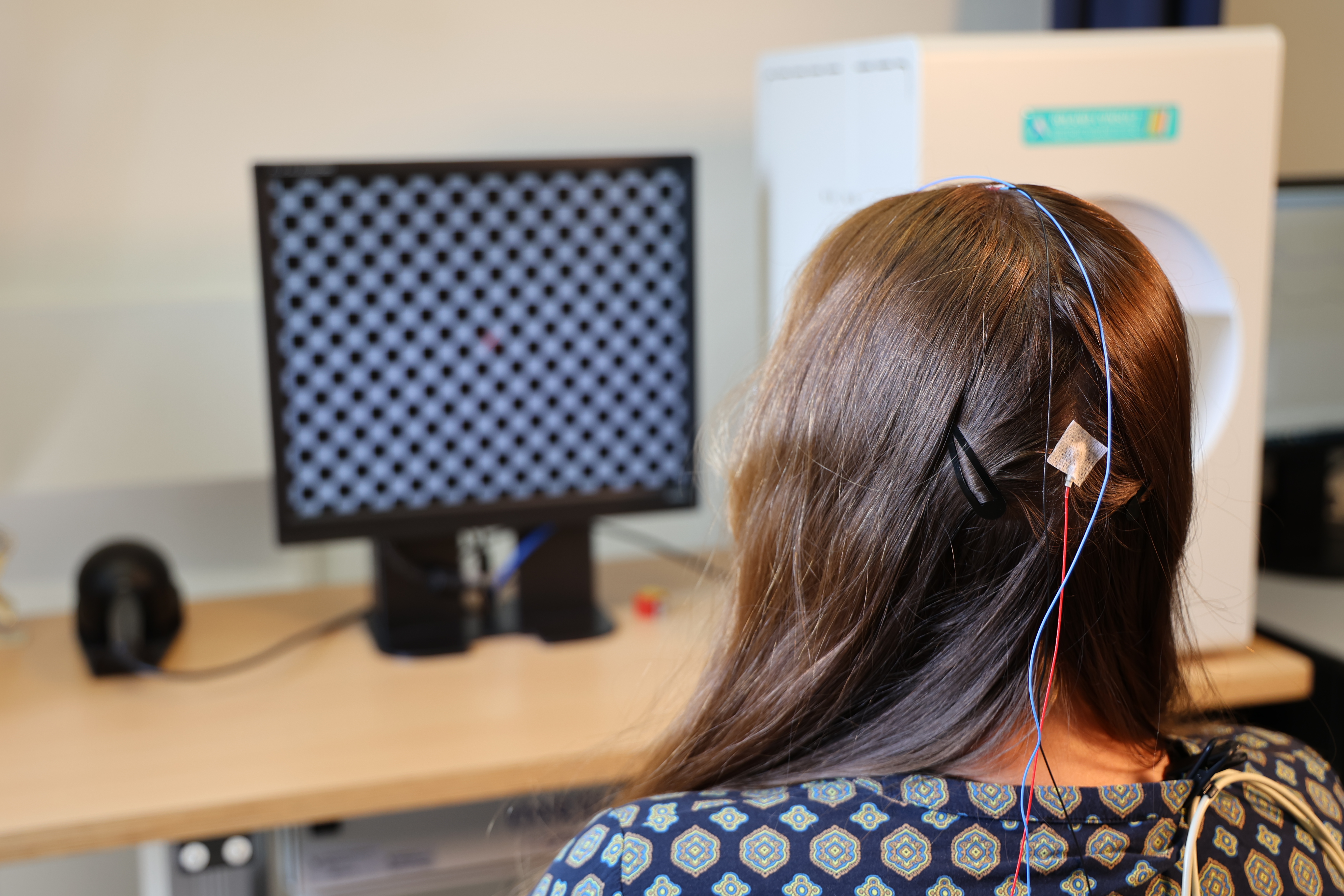
To test the function of the optic nerve, so-called VEP (Visual Evoked Potentials) examinations are performed. Electrodes are taped to the forehead and skull to measure the response of the visual cortex, located in the hindbrain, to light stimuli. The pattern VEP uses checkerboard stimuli, while the visual VEP uses stripe patterns. These examinations are performed, for example, in cases of unclear visual loss or suspected inflammation of the optic nerve, and last about 30 min.
Psychophysical examinations
In psychophysical examinations, the patient says what he or she sees: these are therefore subjective examinations that do not require electrodes.
In dark-adaptation, the adaptation speed from bright light conditions to darkness is examined with an adaptometer. The examination takes about 40 min.
Anomaloscopy allows to determine color vision disorders. This examination can be done for example before choosing a profession where normal color perception is important (e.g. electrical professions).
Mesoptometry (also called nyctometry) allows the determination of twilight vision and glare sensitivity and is performed, for example, for expert opinions regarding fitness to drive.